The smear
Unit 0 of 8
Read the smear itself: what an aspirate is, what each stain shows, how big things are at this scale, and what makes a smear adequate.
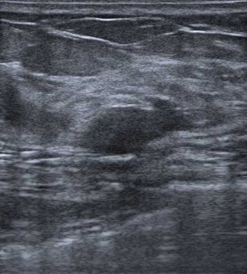
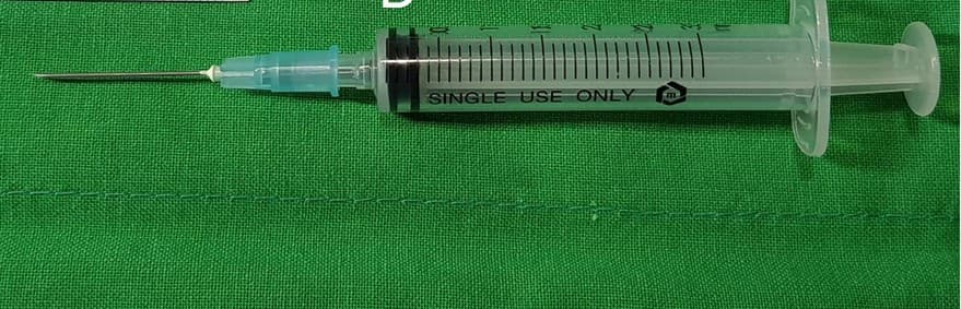
A fine needle aspirate takes a few cells from a breast mass through a thin needle. The cells go onto a glass slide, the slide is smeared and stained, and a scanner photographs it. Every patch in this course is a crop of one of those scans.
Two stains carry the course. Papanicolaou shows nuclear detail and chromatin. May-Grunwald-Giemsa shows cytoplasm, stroma, mucin and the background. The same lesion looks different under each, which is why some patients here have one slide of each.
Before anything is read, the smear has to be worth reading. That is what adequacy means, and the rule is quoted below.
Adequacy, quoted
Adequacy
Seven tissue fragments each consisting of 20 or more epithelial cells to allow assessment of the architecture and the presence or absence of myoepithelial cells, should be a measure of adequacy.
If there is sufficient material on the slides to reach a diagnosis the FNAB is categorized based on that material.
The exceptions
If there are any atypical epithelial features or necrosis, even if there are fewer than seven tissue fragments, the case should be reported as atypical.
Material may be adequate without epithelial cells if it correlates with the clinical and imaging findings, such as pus consistent with an abscess or a proteinaceous background consistent with cyst contents.
C1 Insufficient or inadequate
The smears are too sparsely cellular or too poorly smeared or fixed to allow a cytomorphological diagnosis.
- Risk of malignancy
- 2.6 to 4.8 percent
- Management
Review clinical and imaging findings; if imaging indeterminate or suspicious, repeat FNAB or proceed to CNB.
A cytologist would look for
seven tissue fragments each consisting of 20 or more epithelial cells
pus consistent with an abscess
proteinaceous background consistent with cyst contents
The worked example
Loading the case library.
About 20 trials, 5 to 8 minutes. You can stop at any point.